Primer raziskave podlubnika v jantarju z nedestruktivno metode mikro CT, ki je uporabna tudi pri analizi fosilov. Cilj raziskave je določitev oziroma opis vrste in paleontološke starosti primerka ter študijska evolucijska primerjava z danes živečimi vrstami. 3D računalniški sken mikro CT se lahko uporabi tudi za izdelavo 3D modela s 3D tiskalnikom.
Example of research on a bark beetle embedded in amber using the non-destructive microfocus X-ray computed tomography (microCT) method which is also widely used in fossil analysis.The aim of this research is to describe the species, to determine the paleontological age of the specimen and to perform an evolutionary study in comparison with the living species today. The results of a X-ray microCT scan can also be used to create a 3D model of the specimen by a 3D printer.
Dr. Lucia Mancini, Matjaž Černila
April 2020
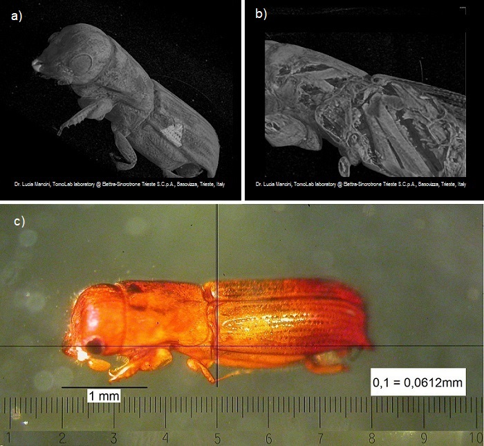
Prostorska upodobitev rentgenskega mikroCT skena podlubnika v jantarju – a) zunanjost primerka in b) navidezni presek 3D slike, ki omogoča vpogled v zgradbo notranjih organov. c) fotografija istega vzorca, posneta z optičnim mikroskopom. (Mikro CT: Lucia Mancini, SYRMEP beamline and TomoLab laboratory of Elettra – Sincrotrone Trieste S.C.p.A.)
Volume renderings obtained by a X-ray microCT scan of a bark beetle in amber – a) exterior of the specimen and b) virtual crop of the 3D image allowing an insight into the structure of the internal organs. c) a photograph of the same specimen taken with an optical microscope.